
What is the difference between MRI and CT? Easily explained
MRI and CT are common diagnostic options in medicine. But what is the difference between the two methods? And what do the abbreviations mean? We explain these points to you in a simple and understandable way.
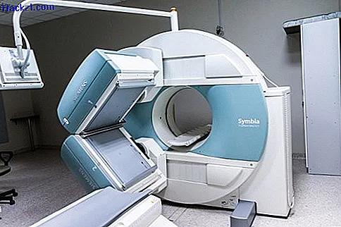
The MRI diagnosis simply explained
Most patients associate an MRI scan with an unpleasant, long-lasting and, above all, noisy scan. Magnetic resonance imaging or magnetic resonance imaging works via a magnetic field. During the 15-20 minute examination, the patient lies in a tube. This is how magnetic resonance therapy works:
- The patient is placed on a couch. This couch is then pushed into the magnetic resonance tomograph. If necessary, a so-called contrast medium is administered to the patient before the examination, which later facilitates the evaluation by the doctors.
- The examination itself takes between 15 and 20 minutes. In the meantime, the patient perceives the tapping noises typical of an MRI scanner.
- The magnetic resonance tomograph builds up a strong magnetic field, approximately 100, 000 times as strong as the earth's magnetic field.
- With this magnetic field, the tomograph "scans" the body and in particular the patient's organs layer by layer.
- The diagnostic option MRT is used by doctors in particular when organs are to be examined.
- According to experts, one advantage of this technology is that, unlike CT examinations, no harmful X-rays are used.
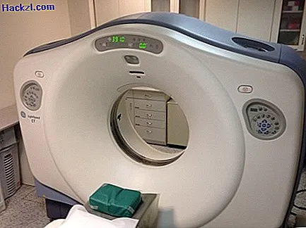
The CT method simply explained
Compared to MRI diagnostics, computed tomography, or CT for short, works with X-rays. During the examination, the patient is also on a couch and in a device. However, the examination time is much shorter.
- Contrast agents are also sometimes used in CT examinations. After administration, the patient lies down on a couch that is pushed into the computer tomograph.
- The device now carries out the examination using X-ray technology. The difference to conventional X-ray examinations is that the whole body is "scanned" during CT.
- During the examination, which usually lasts only a few minutes, the tomograph rotates around the patient to capture every body view.
- The recorded cross-sectional images are now computer-aided assembled into a 3D image.
- Computer tomography offers the advantage of a quick examination and is therefore often used in emergency medicine. In addition, this form of diagnosis is particularly suitable for displaying the human skeleton.
- The disadvantage of CT is the use of X-rays, which can sometimes be harmful to the human organism.
In the next article, you will learn how X-rays work.